Anatomy Of The Back Internal Organs : Female Anatomy Internal Organs Rear Photograph By Leonello Calvetti / New users enjoy 60% off.. Before excreting body waste as urine, the kidneys absorb essential nutrients and electrolytes. The vertebral column (spine) is the bony core of the back. A simple video to introduce the most important organs of the body in english and locate their positions in the body. Internal organs function of the gallbladder in the digestive system. The left kidney, small intestine and descending colon are all found at the lower left side of the back, also known as the left lumbar region.
Schematic drawings of muscles show origins and insertions as vector strands that have the. The spleen is the largest filtering organ in the body and is broken up into two components: The normal arrangement of internal organs is known as situs solitus.although cardiac problems are more common, many people with situs inversus have no medical symptoms or complications resulting from the condition, and. The vertebral column (spine) is the bony core of the back. Lower back pain can be caused by problems with the internal organs.
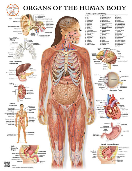
5vndv018mvxyjm from cdn11.bigcommerce.com Situs inversus (also called situs transversus or oppositus) is a congenital condition in which the major visceral organs are reversed or mirrored from their normal positions. The abdomen contains all the digestive organs, including the stomach,. Lower back pain can be caused by problems with the internal organs. Human anatomy female human anatomy picture human anatomy chart human anatomy and physiology body anatomy organs human body organs human body parts human organ diagram. Download 86 internal organs back view stock illustrations, vectors & clipart for free or amazingly low rates! This article looks at female body parts and their functions, and it provides an interactive diagram. Schematic drawings of muscles show origins and insertions as vector strands that have the. Female anatomy of internal organs with skeleton, rear and front views.
They are hard, rigid, and are made up of calcium and phosphorous.
Human anatomy female human anatomy picture human anatomy chart human anatomy and physiology body anatomy organs human body organs human body parts human organ diagram. The internal carotid arteries branch into the skull and circulate blood to the front part of the brain. The back consists of the spine, spinal cord, muscles, ligaments, and nerves. Write on/ wipe off surface. The normal arrangement of internal organs is known as situs solitus.although cardiac problems are more common, many people with situs inversus have no medical symptoms or complications resulting from the condition, and. Anatomy of spinal cord 12 photos of the anatomy of spinal cord anatomy of brain and spinal cord ppt, anatomy of spinal cord tracts, anatomy of the spinal cord exercise 15, development of spinal cord anatomy, overview of spinal cord injury anatomy & physiology, human anatomy, anatomy of brain and spinal cord ppt, anatomy of … The red pulp and the white pulp. When blood passes through the spleen, the red pulp scans the blood for dead blood cells and removes them. This diagram depicts anatomy female 1024×1111 with parts and labels. The organs of the senses and the common integument xi. New users enjoy 60% off. Download 86 internal organs back view stock illustrations, vectors & clipart for free or amazingly low rates! One extremely important part of a dog's skeletal anatomy is the skull.
Write on/ wipe off surface. It is freely movable, especially its distal segment—the hand, which is adapted for grasping and manipulating the. Related posts of anatomy of the back organs anatomy of spinal cord. The organs of the senses and the common integument xi. The white pulp creates new blood cells and places them back into the blood.
Transparent Human Body With Internal Organs Nervous System Lymphatic System And Circulatory System Stock Photo Dissolve from cdn8.dissolve.com Find the perfect human anatomy organs back view stock illustrations from getty images. The normal arrangement of internal organs is known as situs solitus.although cardiac problems are more common, many people with situs inversus have no medical symptoms or complications resulting from the condition, and. The red pulp and the white pulp. They are hard, rigid, and are made up of calcium and phosphorous. When blood passes through the spleen, the red pulp scans the blood for dead blood cells and removes them. The following diagram and paragraphs explain the skeletal anatomy of a dog. Pain may start following inflammation or irritation of an internal organ, or may be a sign of infection. One extremely important part of a dog's skeletal anatomy is the skull.
The vertebral column (spine) is the bony core of the back.
Write on/ wipe off surface. Before excreting body waste as urine, the kidneys absorb essential nutrients and electrolytes. It forms the axial skeleton together with the skull and rib cage. Praise for the thieme atlas of anatomy: Female anatomy of internal organs with skeleton, rear and front views. Lower back pain can be caused by problems with the internal organs. See human back anatomy stock video clips. The internal carotid arteries branch into the skull and circulate blood to the front part of the brain. As with all living beings, the bones surround and protect the internal organs of the body from injury. A simple video to introduce the most important organs of the body in english and locate their positions in the body. The spleen is the largest filtering organ in the body and is broken up into two components: They are hard, rigid, and are made up of calcium and phosphorous. This diagram depicts human heart anatomy for kids 744×991 with parts and labels.
This diagram depicts anatomy female 1024×1111 with parts and labels. Related posts of anatomy of the back organs anatomy of spinal cord. The vertebral arteries follow the spinal column into the skull, where they join together at the brainstem and form the basilar artery , which supplies blood to the rear portions of the brain. Illustration about 3d render of the internal organs as seen from the back, with a silhouette of the body. The spleen is the largest filtering organ in the body and is broken up into two components:
Anatomical Overlays Of The Torso Backside Canvas Print Barewalls Posters Prints Bwc5672625 from images.barewalls.com This diagram depicts human heart anatomy for kids 744×991 with parts and labels. The left kidney, small intestine and descending colon are all found at the lower left side of the back, also known as the left lumbar region. The spleen is the largest filtering organ in the body and is broken up into two components: They are hard, rigid, and are made up of calcium and phosphorous. When blood passes through the spleen, the red pulp scans the blood for dead blood cells and removes them. Human organ diagram back and front view above shows you the unlabeled version of the 3d organ diagram.the following diagrams will show you a much more detailed overview of the location and functions of the major internal organs of the body. See human back anatomy stock video clips. As with all living beings, the bones surround and protect the internal organs of the body from injury.
The spleen is the largest filtering organ in the body and is broken up into two components:
Before excreting body waste as urine, the kidneys absorb essential nutrients and electrolytes. Related posts of anatomy of the back organs anatomy of spinal cord. It is freely movable, especially its distal segment—the hand, which is adapted for grasping and manipulating the. See human back anatomy stock video clips. The red pulp and the white pulp. Schematic drawings of muscles show origins and insertions as vector strands that have the. The normal arrangement of internal organs is known as situs solitus.although cardiac problems are more common, many people with situs inversus have no medical symptoms or complications resulting from the condition, and. The diaphragm forms the upper surface of the abdomen. Learn about 7 conditions that result in symptoms of back pain. Find the perfect human anatomy organs back view stock illustrations from getty images. When blood passes through the spleen, the red pulp scans the blood for dead blood cells and removes them. The internal carotid arteries branch into the skull and circulate blood to the front part of the brain. Human body anatomy female female anatomy muscle shoulder blade pain anatomy back muscles bones man female anatomy body muscles in a body female anatomy muscole shoulder concept muscular sysyem.
It is freely movable, especially its distal segment—the hand, which is adapted for grasping and manipulating the anatomy of back organs. Nervous system , skeleton , front view of muscles , back view of muscles
Anatomy Of The Back Internal Organs : Female Anatomy Internal Organs Rear Photograph By Leonello Calvetti / New users enjoy 60% off.
Reviewed by Creative Ideas
on
June 05, 2021
Rating: 5


Post a Comment